Spina Bifida
What is Spina Bifida
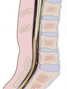
Spina bifida literally means “split or open spine.” It is a congenital malformation that happens in the fetus when there is a kind of cleft, or split, in the back part of the vertebrae.
There are different types of Spina Bifida:
Spina Bifida Occulta

It is the mildest and most common form. It is also called “hidden Spina Bifida” for different reasons. “Occulta” means “hidden” for the fact that is a malformation that is not visible but covered by a layer of skin and also for the fact that about 15% of people have it without knowing it. Spina bifida occulta is usually discovered accidentally when people has an X-ray of their back.
This form usually does not have symptoms and does not cause disability, even if sometimes pain and neurological symptoms may occur.
Spina bifida aperta (“aperta” means “open” in Latin) which includes two other types:

Meningocele
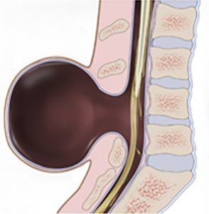
Meningocele is a sac of spinal fluid that protrudes from the spinal column. This fluid does not contain neural tissue so there is usually no nerve damage. It is the least common type of spina bifida and it may cause minor disabilities in children such as difficulty in moving certain muscles, or in controlling certain functions.
Myelomeningocele

This is the most severe types of spina bifida.
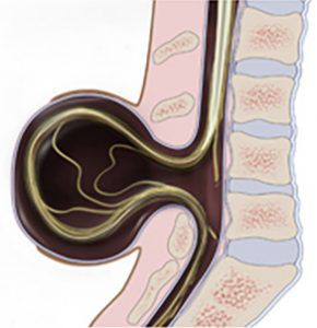
It happens when the bones of the spines do not form in the correct way and a sac with spinal fluid and tissues extends out of the baby’s back. In this case the sac may contain parts of the spinal cord and nerves.
Babies with this condition usually has severe disabilities such as paralysis and other muscle or bone problems. 70 to 90% of children affected by Myelomeningocele have hydrocephalus.
Spina Bifida Diagnosis
There are some prenatal tests that can allow parents to find out this condition in their child.
- The alpha-fetoprotein (AFP) blood test
- Amniocentesis
- Prenatal ultrasound can allow doctors to see most cases of spina bifida aperta
Spina bifida causes
The causes of Spina Bifida are largely unknown. It may be related to environmental and genetic factors and also low levels of the vitamin folic acid during pregnancy are linked to this condition.
Is spina bifida hereditary?
Some evidence suggests that genetic may play a role even if most babies affected by spina bifida have no family history of this condition.
Spina Bifida Treatment
Treatment depends on the severity of the condition that can involve nervous and skeletal systems.
The baby affected by Spina Bifida may undergo a series of surgeries, especially in the first days after birth.
Meningocele requires surgery to put the meninges back into the body and close the hole in the vertebrae.
Myelomeningocele also involves surgery in the very first days after birth to minimize the risk of infection and protect nerve endings and central nervous system.
Sometimes, if myelomeningocele is detected during pregnancy, it is possible to perform a prenatal surgery before the 26th week of pregnancy.
Spina bifida life expectancy, outlook and complications
Babies with meningocele or myelomeningocele may need long-term care from a multispecialty team of surgeons and physicians experts in the field. Depending on the severity of the condition babies may have walking and mobility problems so they may need walking aids, braces, walkers or wheelchair. Other disorders that may be associated with spina bifida can affect the urinary and gastro-intestinal systems, cause mental disabilities and latex allergy.
The aim of the therapy and of doctors is always to help the child become as independent as possible and self-sufficient in daily activities.
Why choose Prof. Portinaro
Prof. Portinaro has a wide experience and particular inclination for neurological conditions.
He performed more than 2,000 orthopedic surgeries on children affected by Spina Bifida and he is also founder and scientific director of a non-profit organization that supports families with children affected by neuromotor disabilities: Fondazione Ariel.
In cases of chronic hip dislocation, Prof. Portinaro’s team also performs hip replacement surgery using the anterior approach on patients of all ages, thanks to the innovative AMIS technique (Anterior Mini Invasive Surgery). This is a minimally invasive anterior hip replacement surgery, a safe, quick procedure (35 minutes) with a fast recovery time (patients can return home independently after just 3 days, without the need for transfusions or drains).
Prof. Portinaro and his team spend a large part of their work in international research projects to discover new treatments for childhood neuromotor disorders.
